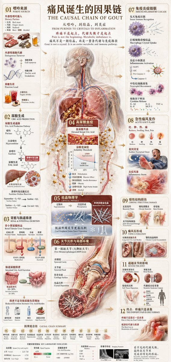
Diabetes Causal Chain Infographic
views183,800
likes509
comments36
shares66
bookmarks370
quotes10
Translated
{"type":"medical infographic poster","topic":"diabetes cause-and-effect chain","language":"Chinese with English subtitles","style":"high-detail biomedical illustration, editorial infographic, warm beige parchment background, realistic anatomy mixed with 3D cutaway organs and molecule diagrams","format":"vertical poster","title":{"main":"The Causal Chain of Diabetes Development","subtitle":"THE CAUSAL CHAIN OF DIABETES","tagline":"From insulin failure to hyperglycemia to systemic damage"},"central_visual":{"description":"large semi-transparent full-body human anatomy in frontal view occupying the center, showing brain, blood vessels, lungs, heart, liver, pancreas, stomach, intestines, kidneys, and surrounding glucose and insulin signaling graphics","emphasis":"metabolic pathways spreading from pancreas and liver through blood circulation to multiple organs"},"layout":{"sections":[{"title":"01 Glucose Enters Life","position":"top-left","count":1,"labels":["GLUCOSE ENTERS LIFE"]},{"title":"02 Pancreas and Insulin","position":"upper-left","count":1,"labels":["PANCREAS & INSULIN"]},{"title":"03 Normal Insulin Action","position":"left-mid","count":1,"labels":["NORMAL INSULIN ACTION"]},{"title":"04 Insulin Resistance: Type 2 Pathway Begins","position":"lower-left","count":1,"labels":["TYPE 2 PATHWAY BEGINS"]},{"title":"05 Liver Keeps Releasing Glucose","position":"top-right","count":1,"labels":["LIVER KEEPS RELEASING GLUCOSE"]},{"title":"06 Beta Cell Failure: Compensation to Failure","position":"center-lower","count":1,"labels":["BETA CELL FAILURE: COMPENSATION TO FAILURE"]},{"title":"07 Type 1 Diabetes Branch","position":"lower-left-center","count":1,"labels":["TYPE 1 DIABETES BRANCH"]},{"title":"08 Hyperglycemia and Blood Chemistry","position":"upper-right-mid","count":1,"labels":["HYPERGLYCEMIA & BLOOD CHEMISTRY"]},{"title":"09 Hyperglycemia Causes Tissue Damage","position":"lower-mid-left","count":1,"labels":["HYPERGLYCEMIA CAUSES TISSUE DAMAGE"]},{"title":"10 Immediate Metabolic Consequences","position":"bottom-mid-left","count":1,"labels":["IMMEDIATE METABOLIC CONSEQUENCES"]},{"title":"11 Microvascular Complications","position":"mid-right-lower","count":1,"labels":["MICROVASCULAR COMPLICATIONS"]},{"title":"12 Macrovascular and Tissue Damage","position":"lower-right","count":1,"labels":["MACROVASCULAR & TISSUE DAMAGE"]},{"title":"13 Long-Term Organ Impact","position":"bottom-right","count":1,"labels":["LONG-TERM ORGAN IMPACT"]},{"title":"14 Diabetes Is Failure of Metabolic Regulation","position":"bottom-left-wide","count":1,"labels":["DIABETES IS FAILURE OF METABOLIC REGULATION"]}],"connectors":"all sections linked by arrows, branching flow lines, circular callouts, and cause-to-effect diagrams leading downward and across the page"},"illustrations":{"organs_count":12,"organs":["digestive tract cross-section with stomach and intestines","pancreas close-up","pancreatic islet microscopic inset","muscle tissue inset","liver close-up","brain in the central body","heart in the chest","intestines in abdomen","retina/eye fundus image","kidney image","nerve/neuropathy image","diseased foot and lower leg vascular damage"]},"small_panels":{"count":6,"items":["glucose and fructose food intake scene with fruit and sweets","insulin receptor signaling diagram with GLUT4 transport","insulin resistance membrane diagram","beta cell decline timeline chart","glucose variability line graph across 24 hours","AGEs/oxidative stress/inflammation tissue damage sequence"]},"photo_style_insets":{"count":8,"items":["sedentary lifestyle couch scene","visceral fat abdominal scan image","glucose strip or glucose measurement tools","obesity/back-fat body photo","sleep disruption tired person","medication bottles or insulin vials","retinopathy eye photo","ulcerated diabetic foot photos"]},"color_palette":{"background":"aged paper beige","primary":"dark brown typography","accent":"red blood vessels and glucose particles","secondary":"orange pancreas and liver tones","tertiary":"blue signaling molecules and cool anatomical highlights"},"typography":{"headline":"large bold Chinese serif-style title with centered English subtitle beneath","section_numbers":"large two-digit dark blocks from 01 to 14","body_text":"dense bilingual explanatory text in compact columns with bullet points and mini-labels"},"composition":"extremely information-dense scientific poster with balanced left-right sidebars, the human body centered, multiple magnified organ cutaways, molecular chains floating around, and a polished magazine-quality educational design","rendering":"ultra-detailed, crisp print-ready poster, realistic anatomy, elegant infographic spacing, subtle parchment texture, soft vignetting, high resolution"}
Machine-translated — the original is what produced this result.


